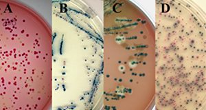
Shiga toxin is a protein found within the genome of a type of virus called a bacteriophage. These bacteriophages can integrate into the genomes of the bacterium E. Coli. Even though most E. coli are benign or even beneficial members of our gut microbial communities, strains carrying Shiga-toxin encoding genes are highly pathogenic in humans and other animals. This six-page fact sheet discusses the two types of Shiga toxins and the best approaches to identifying and determining which Shiga toxin is present. Written by William J. Zaragoza, Max Teplitski, and Clifton K. Fagerquist and published by the Department of Soil and Water Sciences.
http://edis.ifas.ufl.edu/ss654