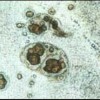
 After many years of diagnostics at the University of Florida and at other laboratories around the country, it appears that Cryptobia iubilans is not uncommon among cichlids, and that environmental and other factors determine the extent of disease.This 3-page fact sheet was written by Ruth Francis-Floyd and Roy Yanong, and published by the UF Department of Fisheries and Aquatic Sciences, September 2014.
http://edis.ifas.ufl.edu/vm077